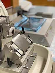
Tissue Processing
Human and/or animal tissue is processed on an automated tissue processor. We are able to process both small biopsy specimens (<0.5cm in length and <0.2cm thick) and larger tissue sections (up to 2cm wide x 3cm long x 0.2 cm thick).
First, tissue is placed in 10% neutral buffered formalin for 90 minutes for large specimens and 20 minutes for biopsies. Then, the tissue is passed through graded alcohols into xylene and paraffin. After the tissue has completed the final paraffin step it is ready for embedding.
- Automated tissue processing of biopsies and large tissue specimens
- Embedding of processed tissue
- Tissue Microarray (TMA) creation
Histology Services
- Embedding of tissue specimen: Tissue is embedded into paraffin cassettes
- Sectioning tissue specimen: We provide the following tissue sectioning.
- Routine: Single section per slide cut at 5 microns.
- Serial sections: Up to three adjacent sections per slide cut at 5 microns.
- Levels: Up to three 5 micron sections per slide. The technician will cut 10-50 microns deeper into the block in between each section depending on what is specified by the researcher.
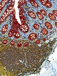
- Hematoxylin and eosin staining: Routine stain for basic tissue morphology performed on automated stainer.
- Unstained Slides: We can provide unstained tissue cut at 5 microns on charged slides for you to stain or process in your own lab.
- Tissue Scrolls: Tissue scrolls can be cut at varying thicknesses and collected in eppindorf tubes for molecular or other testing.
Special Stains
- AFB: Demonstrates Mycobacterium and other acid fast organisms in formalin-fixed, paraffin-embedded tissue.
- Congo Red: Demonstrates amyloid in formalin-fixed, paraffin-embedded tissue. Amyloid will stain pink to red with an apple green birefringence under polarized light.
- Elastic: Demonstrates elastic fibers in formalin-fixed, paraffin-embedded tissue.
- GMS: Demonstrates polysaccharides in the cell walls of fungi and other opportunistic organisms in formalin-fixed, paraffin-embedded tissue.
- Retic: Demonstrates reticular fibers in formalin-fixed, paraffin-embedded tissue.
- Iron: Demonstrates iron in formalin-fixed, paraffin-embedded tissue.
- Jones: Demonstrates carbohydrate components of reticular fibers and basement membranes in formalin-fixed, paraffin-embedded tissue.
- PAS: Demonstrates glycogen, positive reticular fibers, basement membranes, fungus, and neutral mucopolysaccharides in formalin-fixed, paraffin-embedded tissue.
- Diastase: Used in conjunction with PAS to aid in the demonstration of glycogen in formalin-fixed, paraffin-embedded tissue.
- Trichrome: Demonstrates connective tissue, muscle, and collagen fibers in formalin-fixed, paraffin-embedded tissue.
Immunohistochemistry
- Single stain IHC
- We are able to stain formalin-fixed, paraffin-embedded tissue cut at 5 microns with a vast array of immunohistochemical stains. You may choose from the list of antibodies we have already worked up, or provide your own antibody and control for us to work up for you. A list of all of our current antibodies can be found here.
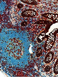
- Multiplex (up to 6 antibodies)
- We have several different chromogens which allows us to stain up to 5 different antibodies on one slide. You may choose from the list of antibodies we have already worked up, or you may provide your own. Researcher provided antibodies can be from mouse, rat, rabbit, or goat.